Nervous Coordination
Coordination is the process of harmonizing actions in the body to achieve specific goals. The nervous system in mammals is divided into two main parts:
- Central Nervous System (CNS): This includes the brain, protected by the cranium, and the spinal cord, located in the vertebral column.
- Peripheral Nervous System (PNS): Links the CNS to the body’s receptors and effectors.
The Brain
The brain is a highly specialized organ composed of millions of nerve cells (neurons). It is protected by the skull and is divided into three main parts:
-
Forebrain: Includes the cerebrum,
olfactory lobes, thalamus, and hypothalamus.
-
Cerebrum: The largest brain
part, consisting of right and left
hemispheres.
- Functions: Controls intelligence, learning, memory, imagination, reasoning, voluntary actions, and sensory interpretation.
-
Olfactory Lobes: Small
structures at the front of the brain.
- Functions: Receives sensory impulses for smell.
-
Thalamus: Two ovoid
structures at the back of the forebrain.
- Functions: Processes sensations and maintains consciousness.
-
Hypothalamus: Located below
the thalamus.
- Functions: Controls sleep, appetite, body temperature, and alertness.
-
Cerebrum: The largest brain
part, consisting of right and left
hemispheres.
-
Midbrain: Connects the forebrain
and hindbrain.
- Functions: Handles vision (via optic lobes) and transmits impulses between the forebrain and hindbrain.
-
Hindbrain: Includes the cerebellum,
medulla oblongata, and pons varolii.
-
Cerebellum: Oval-shaped,
located below the cerebrum, and less
convoluted.
- Functions: Maintains balance, coordinates muscle actions, and processes impulses from the ears and skin.
- Pons Varolii: Connects the cerebellar hemispheres.
-
Medulla Oblongata: The
posterior part of the brain that continues
into the spinal cord.
- Functions: Regulates involuntary actions like respiration, heartbeat, digestion, and blood pressure.
-
Cerebellum: Oval-shaped,
located below the cerebrum, and less
convoluted.
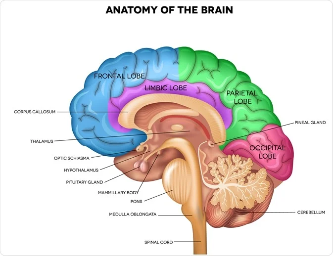
The Spinal Cord
The spinal cord extends from the medulla oblongata and is protected by the backbone and three meninges layers. It has an outer white matter (axon bundles) and an inner grey matter (neurons).
Functions:
- Coordinates reflex actions like knee-jerk and sweating.
- Acts as a pathway for communication between the brain and the body.
Peripheral Nervous System (PNS)
The PNS connects the CNS to sensory organs and effectors. It consists of:
- 12 pairs of cranial nerves.
- 31 pairs of spinal nerves.
The PNS is divided into:
- Somatic Nervous System (SNS): Handles voluntary actions and responses to external stimuli.
-
Autonomic Nervous System (ANS):
Regulates involuntary actions like heartbeat and
digestion. It has two parts:
- Sympathetic Nervous System: Prepares the body for action during stress.
- Parasympathetic Nervous System: Promotes rest by slowing the heartbeat and relaxing muscles.
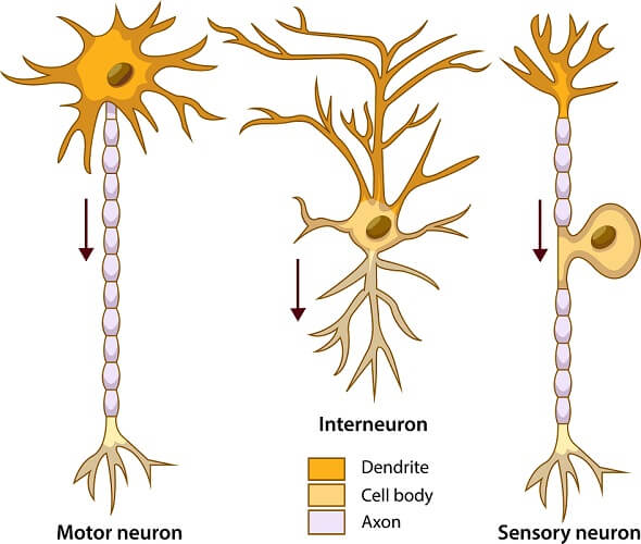
Neurons
Neurons are the basic units of the nervous system. They transmit electrical impulses and consist of:
- Cell Body: Contains the nucleus and cytoplasm.
- Dendrites: Carry impulses toward the cell body.
- Axon: Transmits impulses away from the cell body, insulated by a myelin sheath.
Types of Neurons:
- Sensory Neurons: Transmit impulses from receptors to the CNS.
- Motor Neurons: Carry impulses from the CNS to effectors (e.g., muscles).
- Intermediate Neurons: Connect sensory and motor neurons within the CNS.
Impulse Transmission
Impulse transmission involves three phases:
- Resting Potential: No impulse; the cell is polarized with more sodium ions outside and potassium ions inside.
- Action Potential: The nerve is depolarized as sodium ions enter and potassium ions exit.
- Repolarization: Resting potential is restored after impulse transmission.
Impulses are transmitted chemically at synapses using neurotransmitters like acetylcholine.
Reflex and Voluntary Actions
Reflex Actions: Involuntary responses to stimuli, like sneezing and blinking, involving a reflex arc of sensory and motor neurons.
Voluntary Actions: Conscious, deliberate activities such as writing, walking, and dancing.
Conditioned Reflexes
Conditioned reflexes are learned responses, like typing or swimming, acquired through practice and repetition.
The concept of the conditioned reflex was first demonstrated by a Russian scientist, Pavlov, in 1910. He observed that a dog naturally salivates when food is presented. Pavlov altered this natural response by ringing a bell just before presenting food to the dog. After repeating this sequence several times, he rang the bell without providing any food. Remarkably, the dog still salivated upon hearing the bell. This experiment showed that, in a conditioned reflex, the stimulus (e.g., the bell) and the response (e.g., salivation) do not need to be directly related, as the bell had no inherent connection to the food.